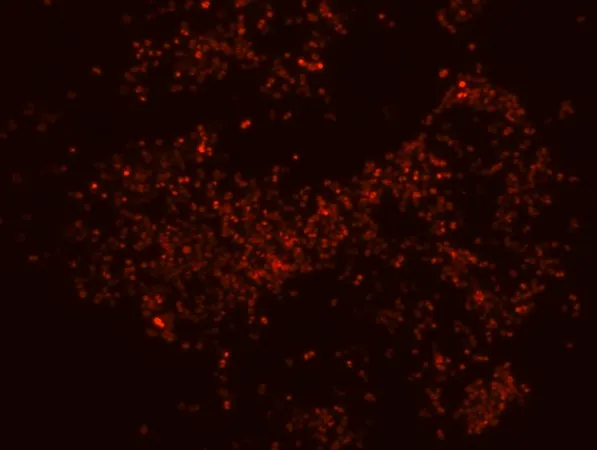
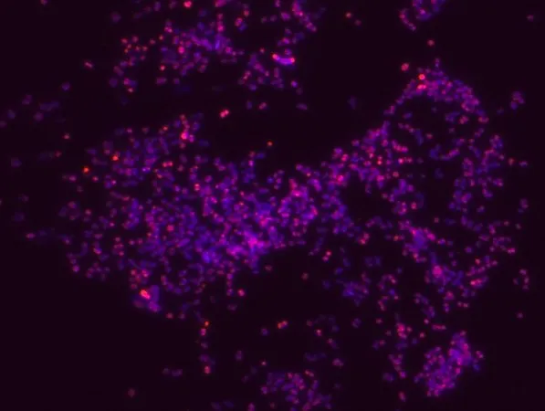

| 货号 | 包装规格 | 操作 |
|---|---|---|
| MBD0029-50UL | 50 μL |
Fluorescent In Situ Hybridization technique (FISH) is based on the hybridization of fluorescent labeled oligonucleotide probe to a specific complementary DNA or RNA
sequence in whole and intact cells.1 Microbial FISH allows the visualization, identification and isolation of bacteria due to recognition of ribosomal RNA also in unculturable samples.2
FISH technique can serve as a powerful tool in the microbiome research field by allowing the observation of native microbial populations in diverse microbiome environments, such as samples from human origin (blood3 and tissue4), microbial ecology (solid biofilms5 and aquatic systems6) and plants7. It is strongly recommended to include positive and negative controls in FISH assays to ensure specific binding of the probe of interest and appropriate protocol conditions. We offer positive (MBD0032/33) and negative control (MBD0034/35) probes, that accompany the specific probe of interest.
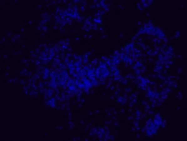
Porphyromonas gingivalis probe specifically recognizes P. gingivalis cells. P. gingivalis, is a gram negative bacterium which is an etiologic agent of adult periodontitis, a chronic inflammatory disease characterized by the destruction of the supportive tissue surrounding teeth. Studies have shown that LPS from P. gingivalis plays an important role in this disease.8-11 The association of the oral microbiota, including P. gingivalis, with various pathological states has been reported. These include development of Alzheimer′s disease12, role in oral cancers13, preterm birth14 and rheumatoid arthritis15. FISH technique was successfully used to identify P. gingivalis with the probe in various samples such as pure culture (as described in the figure legends), dental implants16,17 , periapical tooth lesions18, saliva19, brain tissue20, gingival and aortic tissues21, biofilms from voice prostheses22, subgingival biofilm23, aortic wall tissue24, and infected HeLa cells25. Moreover, FISH can be implicated to identify P. gingivalis in tumor tissue26, multispecies biofilm27, multispecies oral biofilms28 and pure culture and buccal epithelial cells29.
荧光原位杂交(FISH)探针,识别牙龈卟啉单胞菌细胞
观察、鉴定和分离牙龈卟啉单胞菌细胞。观察不同微生物群环境中的天然牙龈卟啉单胞菌细胞群。在细菌混合群中特异性、灵敏性和稳健性地鉴定牙龈卟啉单胞菌。特异性、即使在样本中牙龈卟啉单胞菌丰度较低的情况下,也能进行灵敏和稳健的鉴定。FISH可通过避免污染细菌的检测完成基于PCR的检测方法。牙龈卟啉单胞菌的形态学,并允许研究生物膜结构。鉴定临床样品中的牙龈卟啉单胞菌,如肿瘤和脑组织(例如福尔马林固定的石蜡包埋(FFPE)样品)、唾液和口腔以及医疗设备中的牙龈卟啉单胞菌,如牙种植体和声音假体。在牙龈卟啉单胞菌的自然栖息地中检测牙龈卟啉单胞菌的能力是研究宿主-微生物组相互作用的必要工具。
| 英文名称 | Porphyromonas gingivalis FISH probe - Cy3 |
| NACRES | NA.55 |
| UNSPSC | 12352200 |
| 技术 | FISH:适用 |
| 荧光 | λex 550 nm; λem 570 nm(Cy3) |
| 运输状态 | 干冰 |
| 储存温度 | −20°C |
| 质量等级 | 200 |
| 包装规格(货号 MBD0029-50UL) | 50 μL |